Search results
Search for "X-ray diffraction (XRD)" in Full Text gives 336 result(s) in Beilstein Journal of Nanotechnology. Showing first 200.
Controllable physicochemical properties of WOx thin films grown under glancing angle
Beilstein J. Nanotechnol. 2024, 15, 350–359, doi:10.3762/bjnano.15.31

- bias voltage was applied to the p-Si substrates, whereas the WOx films were kept grounded. The crystallinity of the WOx films was examined using X-ray diffraction (XRD) (Bruker) under Bragg–Brentano geometry (θ–2θ) in an angular window of 2θ = 20° to 80°. The chemical composition of the WOx films was


Vinorelbine-loaded multifunctional magnetic nanoparticles as anticancer drug delivery systems: synthesis, characterization, and in vitro release study
Beilstein J. Nanotechnol. 2024, 15, 256–269, doi:10.3762/bjnano.15.24

- -resolution analytical electron microscope (FE-SEM, Thermo Scientific, Apreo 2S LoVac) and a scanning transmission electron microscope (STEM, Phillips XL, 30 ESEM-FEG/EDAX) operating at 120 kV acceleration voltage. The structure of the nanoparticles was analyzed by X-ray diffraction (XRD, PANalytical, Xpert
- between peak broadening and particle size in X-ray analysis. In this equation, the symbols D, K, λ, β, and θ represent the particle size, Scherrer shape factor (here 0.89), X-ray wavelength (0.15418 nm), half-maximum width, and diffraction angle, respectively [43]. Using the X-ray diffraction (XRD


Modification of graphene oxide and its effect on properties of natural rubber/graphene oxide nanocomposites
Beilstein J. Nanotechnol. 2024, 15, 168–179, doi:10.3762/bjnano.15.16

- characterized by X-ray diffraction (XRD), Fourier-transform infrared spectroscopy, contact angle, thermal gravimetric analysis, and scanning electron microscopy. The XRD results showed the appearance of an amorphous region of silica particles at a diffraction angle of 22°. The formation of silica was
- conditions to determine the ideal condition to modify GO for grafting onto NR. The GO-VTES products were characterized using X-ray diffraction (XRD), contact angle, 29Si NMR, Fourier-transform infrared spectroscopy (FTIR), and morphology analysis. The GO-VTES was expected to improve the mechanical properties


Berberine-loaded polylactic acid nanofiber scaffold as a drug delivery system: The relationship between chemical characteristics, drug-release behavior, and antibacterial efficiency
Beilstein J. Nanotechnol. 2024, 15, 71–82, doi:10.3762/bjnano.15.7

- separation during the electrospinning process [17][38][39], leading to the formation of a BBR-rich phase on the surface of nanofibers. The crystallinity of the PLA pellet and electrospun nanofiber scaffolds were examined by X-ray diffraction (XRD) analysis (Figure 3B). The XRD pattern of the PLA pellet shows


Experimental investigation of usage of POE lubricants with Al2O3, graphene or CNT nanoparticles in a refrigeration compressor
Beilstein J. Nanotechnol. 2023, 14, 1041–1058, doi:10.3762/bjnano.14.86

- nanoparticles is presented separately in the subsequent sections to verify the catalog information provided by the manufacturer. In the characterization of the nanoparticles used in the study, field-emission scanning electron microscopy (FE-SEM), energy-dispersive X-ray spectroscopy (EDS), and X-ray diffraction
- (XRD) analyses were performed. Characterization of Al2O3 nanoparticles The morphological features of the Al2O3 nanoparticles were investigated with the help of FE-SEM micrograph (Figure 2a). It is seen that the Al2O3 nanoparticles exhibit amorphous nature. Thus, it can be stated that the particle size


Exploring internal structures and properties of terpolymer fibers via real-space characterizations
Beilstein J. Nanotechnol. 2023, 14, 1004–1017, doi:10.3762/bjnano.14.83

- , this distinction in fundamental chemistry has significant implications for the structures and properties of the resulting fibers. To date, structure–property characterizations of Technora® in the literature have primarily focused on (i) X-ray diffraction (XRD), (ii) nuclear magnetic resonance (NMR


A wearable nanoscale heart sound sensor based on P(VDF-TrFE)/ZnO/GR and its application in cardiac disease detection
Beilstein J. Nanotechnol. 2023, 14, 819–833, doi:10.3762/bjnano.14.67

- underwent characterization through electron microscopy, X-ray diffraction (XRD), and piezoelectric performance testing. The results indicated that the piezoelectric film with a composition ratio of 12% P(VDF-TrFE) + 10% ZnO + 0.1% GR exhibited superior performance regarding various aspects. Consequently, in


Nanostructured lipid carriers containing benznidazole: physicochemical, biopharmaceutical and cellular in vitro studies
Beilstein J. Nanotechnol. 2023, 14, 804–818, doi:10.3762/bjnano.14.66

- medium observed after the initial stage could be attributed to the gradual release of drug molecules from the matrix core, where the drug is mainly located according to X-ray diffraction (XRD) results [33]. Remarkably, although our NLC possess a comparatively lower drug load, the maximal accumulated drug


Silver nanoparticles loaded on lactose/alginate: in situ synthesis, catalytic degradation, and pH-dependent antibacterial activity
Beilstein J. Nanotechnol. 2023, 14, 781–792, doi:10.3762/bjnano.14.64

- potential measurements, which were measured on a nanoPartica Horiba SZ-100 (Japan). Fourier-transform infrared (FTIR) spectra were obtained using a Bruker Tensor 27 FTIR spectrophotometer (Germany). X-ray diffraction (XRD) patterns were collected using a Bruker D8 Advance X-ray diffractometer. The


Titania nanoparticles for photocatalytic degradation of ethanol under simulated solar light
Beilstein J. Nanotechnol. 2023, 14, 616–630, doi:10.3762/bjnano.14.51

- in the second batch of samples, that is, in series “b”. The elemental composition of the TiO2 powders was estimated by EDS performed inside a scanning electron microscope, FEI Quanta Inspect S, at 15 kV in high vacuum. The crystalline structures and phase concentrations were determined from X-ray
- diffraction (XRD) patterns, measured by an X-ray diffractometer Panalytical X’Pert MPD theta–theta, and the morphological properties were determined by transmission electron microscopy (TEM), high-resolution transmission electron microscopy (HRTEM), and selected-area electron diffraction (SAED) measurements


Mixed oxides with corundum-type structure obtained from recycling can seals as paint pigments: color stability
Beilstein J. Nanotechnol. 2023, 14, 467–477, doi:10.3762/bjnano.14.37

- colorimetric stability of the aluminates applied as synthetic inorganic pigments [14]. Characterization The samples were characterized by powder X-ray diffraction (XRD) carried out in a Bruker D2 Phaser Diffractometer (Berlin, Germany) with Cu Kα emission (λ = 1.5418 Å) and equipped with a LynxEye high


Evaluation of electrosynthesized reduced graphene oxide–Ni/Fe/Co-based (oxy)hydroxide catalysts towards the oxygen evolution reaction
Beilstein J. Nanotechnol. 2023, 14, 420–433, doi:10.3762/bjnano.14.34
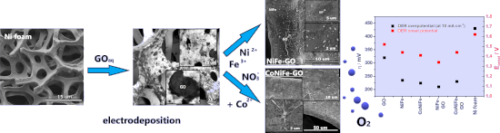
- ]. The spectra were obtained using the total electron yield (TEY) detection mode, which can sample down to a depth of a few nanometers at room temperature. The beamline optics was optimized to perform the experiment with an energy resolution of 200 meV and better. X-ray diffraction (XRD) measurements


Formation of nanoflowers: Au and Ni silicide cores surrounded by SiOx branches
Beilstein J. Nanotechnol. 2023, 14, 133–140, doi:10.3762/bjnano.14.14

- atomic numbers show brighter contrasts. EDS measurements were performed to obtain the element distribution in the target areas. X-ray diffraction (XRD, Siemens D-5000) analyses were conducted in Bragg–Brentano mode using Cu Kα irradiation at 40 kV. The height distribution of the areas of interest was


Liquid phase exfoliation of talc: effect of the medium on flake size and shape
Beilstein J. Nanotechnol. 2023, 14, 68–78, doi:10.3762/bjnano.14.8

- powder was exfoliated in each liquid medium by exposure to mechanical energy provided by an ultrasonic bath (full details in the Experimental section). Talc was manually milled down to a fine powder and characterized by X-ray diffraction (XRD). Figure 1a displays the results. All peaks are assigned to
- obtaining information on thousands of flakes and using appropriate statistical descriptions to analyze the data. Experimental Materials. Talc was obtained through a donation of a sample from Minas Gerais state, Brazil. X-ray diffraction (XRD) was performed to characterize the sample composition. The rock
- measurements were performed on silicon substrates with an oxide layer, Si/SiOx. Substrates were functionalized with (3-aminopropyl)triethoxysilane (APTES) following the procedure reported by Fernandes and co-workers [24]. X-ray diffraction. XRD was performed in a Rigaku Geigerflex 2037 diffractometer with a


Two-step single-reactor synthesis of oleic acid- or undecylenic acid-stabilized magnetic nanoparticles by thermal decomposition
Beilstein J. Nanotechnol. 2023, 14, 11–22, doi:10.3762/bjnano.14.2

- were uniform and the single spots were not visible proved that the crystallites were very small. These results correspond well with data from X-ray diffraction (XRD), according to which the average size of the crystallites for all prepared nanoparticles was 4.5–9 nm. The average crystallite size did


Photoelectrochemical water oxidation over TiO2 nanotubes modified with MoS2 and g-C3N4
Beilstein J. Nanotechnol. 2022, 13, 1541–1550, doi:10.3762/bjnano.13.127

- of materials The morphology, the phase, and the vibrational characteristics of the surface functional groups of the materials were observed by field-emission scanning electron microscopy (FESEM), X-ray diffraction (XRD), and Fourier-transform infrared spectroscopy (FTIR). Diffuse reflectance


A TiO2@MWCNTs nanocomposite photoanode for solar-driven water splitting
Beilstein J. Nanotechnol. 2022, 13, 1520–1530, doi:10.3762/bjnano.13.125

- nanocomposite characterizations The surface morphology of MWCNTs and the TiO2@MWCNTs nanocomposite is characterized by using field-emission scanning electron microscopy (FE-SEM, S4800) and transmission electron microscopy (TEM, JEOL-1400). The crystallization behavior of the catalysts is analyzed by X-ray
- diffraction (XRD, D2 PHASER). The chemical structure of the samples is characterized using Fourier-transform infrared spectroscopy (FTIR, Brucker 27). The electrochemical measurements are carried out on a MPG2 Biologic system with a three-electrode cell controlled by ECLab® software. Diffuse reflectance


In search of cytotoxic selectivity on cancer cells with biogenically synthesized Ag/AgCl nanoparticles
Beilstein J. Nanotechnol. 2022, 13, 1505–1519, doi:10.3762/bjnano.13.124

- pineapple peel extracts and their behavior on the breast cancer cell line MCF-7 is shown. Bioreactions were monitored at different temperatures. Fourier-transform infrared spectroscopy (FTIR), ultraviolet–visible spectroscopy (UV–vis), thermogravimetric analysis (TGA), X-ray diffraction (XRD), energy


Rapid fabrication of MgO@g-C3N4 heterojunctions for photocatalytic nitric oxide removal
Beilstein J. Nanotechnol. 2022, 13, 1141–1154, doi:10.3762/bjnano.13.96

- properties of the materials. Scanning electron microscopy (SEM) and high-resolution transmission electron microscopy (HR-TEM) were used to assess the morphology of the materials. The crystal phase of the materials was determined by X-ray diffraction (XRD) with a measurement range of 10°–80°. Fourier


Green synthesis of zinc oxide nanoparticles toward highly efficient photocatalysis and antibacterial application
Beilstein J. Nanotechnol. 2022, 13, 1108–1119, doi:10.3762/bjnano.13.94

- reactions that form zinc resinate are shown in Equation 1 and Equation 2. The schematic illustration of the synthesis of ZnO nanoparticles is shown in Figure 1. Methods for determining the characterization of the synthesized material The phase of the synthesized material was determined by X-ray diffraction
- (XRD) using a Bruker D8 advanced X-ray diffractometer equipped with Cu Kα radiation (λ = 1.5418 Å). The morphology and size of the synthesized material were determined by field emission scanning electron microscopy (FESEM) on a Hitachi S-4800 at 15 kV and high-resolution transmission electron


Hierarchical Bi2WO6/TiO2-nanotube composites derived from natural cellulose for visible-light photocatalytic treatment of pollutants
Beilstein J. Nanotechnol. 2022, 13, 745–762, doi:10.3762/bjnano.13.66

- the practical content (72.9 wt %) of the Bi2WO6 component in the 70%−Bi2WO6/TiO2-NT nanocomposite. Characterization Powder X-ray diffraction (XRD) patterns of the samples were obtained from the Rigaku Ultima IV diffractometer with a Cu Kα (λ = 0.15405 nm) radiation source. Fourier transform infrared


A nonenzymatic reduced graphene oxide-based nanosensor for parathion
Beilstein J. Nanotechnol. 2022, 13, 730–744, doi:10.3762/bjnano.13.65

- phase of GO and RGO was characterized by X-ray diffraction (XRD) using a X’pertpro MPD XRD (PAN analytical B.V., the Netherlands) with Cu Kα radiation (λ = 1.5406 Å). Scanning electron microscopy (SEM) of the modified electrode was conducted on a JEOLEVO® 18 special edition (model: ZEISS EVO-MA 10) at


Nanoarchitectonics of the cathode to improve the reversibility of Li–O2 batteries
Beilstein J. Nanotechnol. 2022, 13, 689–698, doi:10.3762/bjnano.13.61

- the carbonization process. After the carbonization and chemical etching processes, the sizes of the ZnxCoy particles were slightly decreased due to the thermal evaporation of organic linkers and metal ions, maintaining free spaces in the particles. According to the X-ray diffraction (XRD) patterns of


Sodium doping in brookite TiO2 enhances its photocatalytic activity
Beilstein J. Nanotechnol. 2022, 13, 599–609, doi:10.3762/bjnano.13.52

- bandgap values of these four samples. Structural phase diagram, chemical composition, and morphology The crystal structure of samples calcinated at 300–900 °C was characterized by powder X-ray diffraction (XRD), as shown in Figure 3a. The sample calcinated at 300 °C is a mixture of brookite (B) and


A new method for obtaining the magnetic shape anisotropy directly from electron tomography images
Beilstein J. Nanotechnol. 2022, 13, 590–598, doi:10.3762/bjnano.13.51

- microscopy (STEM) operation mode, using the high-angle annular dark-field (HAADF) detectors and an appropriate camera length. The TEM specimen was prepared by a standard powder method, using a 300 Mesh, lacey Carbon, Cu grid. X-ray diffraction (XRD) has been performed on MNPs using a Bruker D8 Advance


